The sense of sight
Biology / / July 04, 2021
The eyesight us allows us to know the world around us through the images that are projected in our eyes, and that are interpreted in our brain.
The organ in charge of the sense of sight is the eye.
Each of our eyes captures the images of what we have around us, each one with a slight difference in angles, which our brain unifies into a single image; However, this slight difference in the information that our eyes capture also allows us to calculate the depth of the images and calculate the distances. This ability to calculate distances and the depth of objects is called stereoscopy.

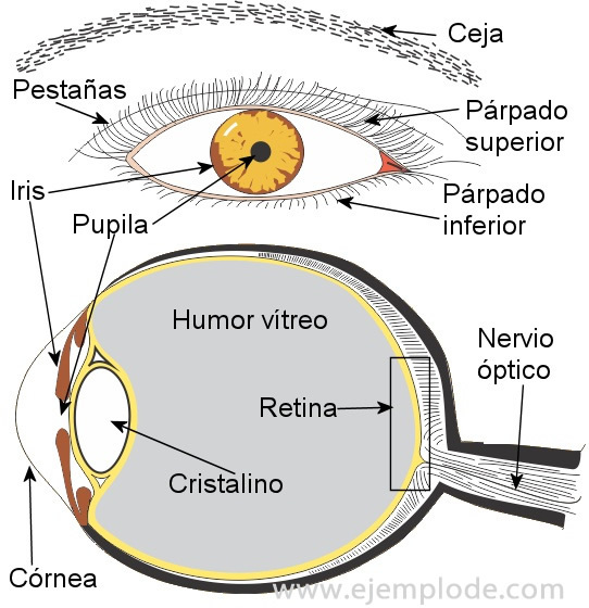
The parts of the eye are:
Eyeball
It is the structure that forms the eye as a whole.
Eyelids
They are covered with skin provided with muscles, which allow them to open and close.
Conjunctiva
It is a membrane that lines the space between the eyeball and the eyelids. It allows to keep the eye moist by being moistened with tears.
Tear sacs
They are two sacs where tears are formed, which serve as a lubricant for the conjunctival membrane, between the eyelids and the eye, helping together with the movement of the eyes. eyelids, so that the eye does not dry out and at the same time that if there is any impurity or some irritant (physical or chemical) it can come out when a greater amount of tears.
Cornea
It is the outermost structure of the eye, which is transparent and shaped like a dome. It has the function of concentrating the light and the image, so that they pass through the pupil.
Iris
It is the part that gives color to our eyes and that surrounds the pupil. It is made up of pigmented cells and small muscles, which react to light, opening if there is little light. (the pupil looks large and the iris decreases) or closing the passage of light (the iris is more visible and the pupil looks little).
Pupil
It is the black point of the eyes, it is where the light enters, and where the image that passes through the cornea is concentrated.
Crystalline
It is a structure that is shaped like a biconvex lens, that is, it is bulky on both sides of the center and thin towards the edges. On its edges it is attached to muscles, which stretch or compress it, causing it to change its curvature. As it passes through the lens, the image perceived through the cornea is inverted and is projected inverted into the eye.
Vitreous humor
The vitreous humor is a gelatinous mass, which under normal conditions is transparent and fulfills the function of transmitting light and images, as well as maintaining the shape of the eye.
Retina
It is the part of the back of the eye, where the image that passes through the lens is formed. It is covered by a layer of nerve receptors called cones and rods, which convert light impulses into nerve signals that are sent to the brain. The rods and cones are not evenly distributed throughout the retina. There is a point approximately in the center of the image, where there are a greater number of receptors, and which is also where the image is perceived with greater clarity. This point is called fovea and it is in the form of a small depression.
Optic nerve
The optic nerve transmits nerve signals from the retina to the brain, where they are recomposed as visual perceptions, and where images are interpreted and stored.
Eye muscles
They are muscles that are inserted into the body of the eye, and they are what allow us to move our eyes to one side, up and down.


