Definition of Cranial Pairs
Miscellanea / / July 04, 2021
By Dra. Maria de Andrade, CMDF 21528, MSDS 55658., on Oct. 2018
 The nervous system carries out the control of the various functions of the body, for this it needs to collect information from both the outside and the internal environment, process it and then send signals that will translate into movements, hormonal secretion, changes in the activity of the viscera or even manifestations motivate and learning.
These functions are carried out in the Central Nervous System, located within the skull and in the spinal canal of the spine. The transport of information and effector signals from the brain and higher centers to the different organs and systems occurs thanks to the processes that form the nerves and that constitute the peripheral nervous system.
This system is made up of the nerves that emerge from the spinal column originating at the level of the spinal cord, which are the nerves spinal, as well as those from the brain and brainstem that are externalized when passing through the different holes in the skull known as pairs cranial.
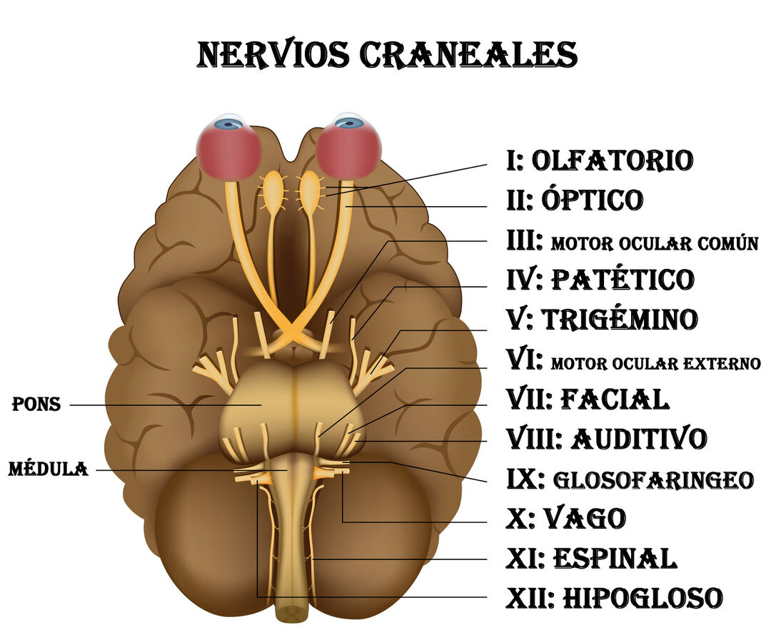
The cranial nerves are paired nerves as their name indicates, since there is one for each side of the body, There are a total of 12 ribs and are named with Roman numerals, as we can see in the art on the side of the text, which you can expand with one click.

Functions of the cranial nerves
Olfactory. It originates in nerve cells related to the sense of smell located in the roof of the nostrils, from there it goes to the higher centers to allow olfaction.
Optical. The visual impulses originate in the cells of the retina (rods and cones) and pass through a system of 4 neurons that end in the optic nerve, responsible for carrying information to the visual cortex located in the occipital lobe of the brain.
Common ocular motor or oculomotor. Controls most of the eye muscles allowing eye movements to be carried out up, down and inwards, it also controls the diameter of the pupil and the accommodation of the lens essential for vision close.
Trochlear. It works by moving the eye up and down.
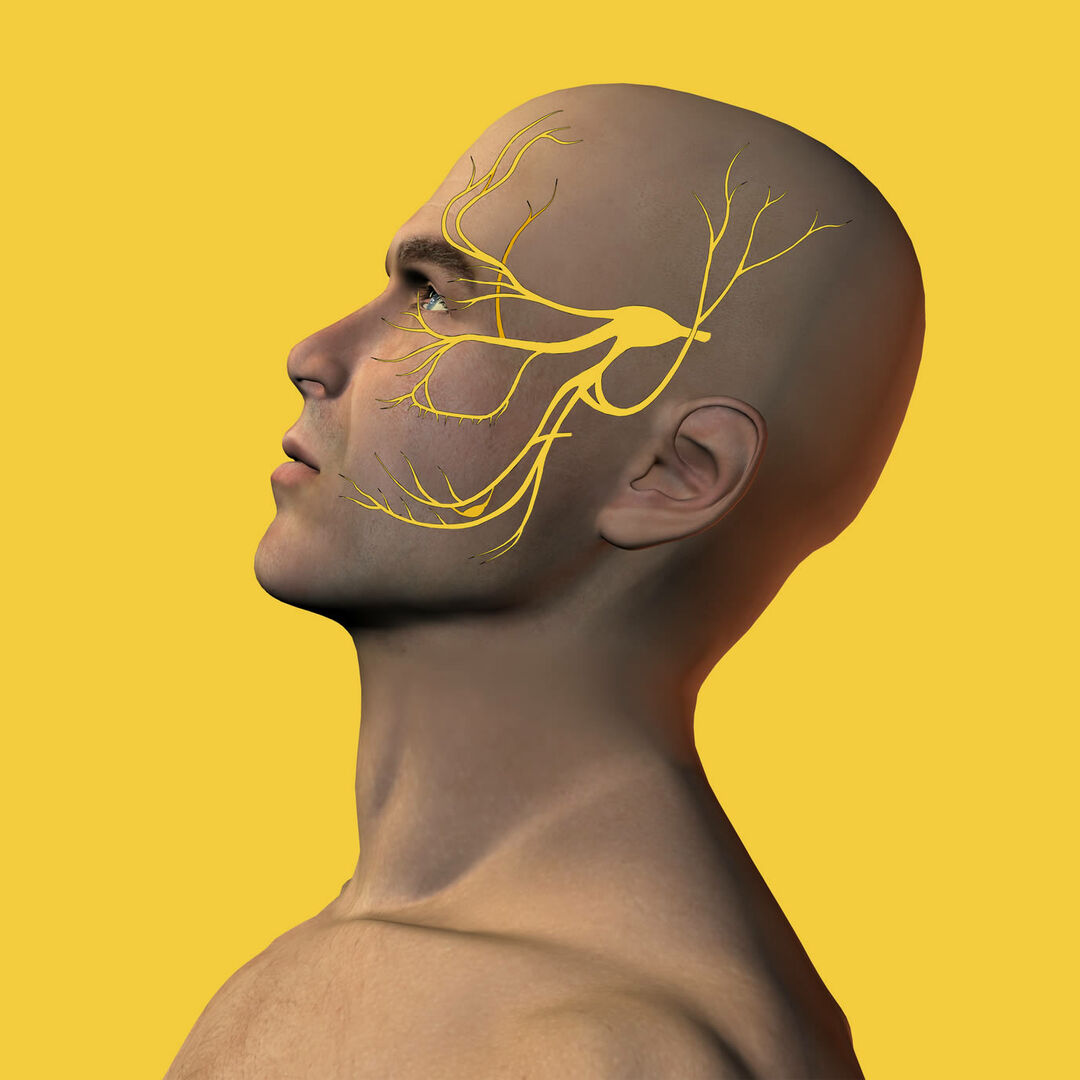
Trigeminal. It is the nerve responsible for the sensitivity of the face and the motor skills of the chewing muscles. It has three branches, upper or ophthalmic, upper middle maxilla and lower lower maxilla.
External ocular motor. It is dedicated exclusively to movements that allow the eye to be drawn outward.
Facial. It is the nerve that allows the mobility of the face and the ear muscles, it also provides the sensitivity of the anterior two thirds of the language and controls functions of the lacrimal and salivary glands.
Vestibulocochlear. It originates in the inner ear, allows to carry the electrical signals originated by the vibrations of the eardrum to the cerebral auditory centers so that the hearing takes place. It consists of a vestibular branch that transmits information about the position of the head in space, as well as its movements, which is essential for the control of the Balance.
Glossopharyngeal. Provides sensitivity to the throat and back of the palate while also controlling its mobility.
Vague. It is the longest cranial nerve. It emerges from the skull and descends to the thorax and abdomen to give sensory and autonomic (parasympathetic) innervation to the organs of the apparatus. cardiovascular, respiratory and digestive, making it an important regulator of functions such as the control of blood pressure, frequency cardiac, breathing, bowel movements and digestion.
Spinal or accessory. It is a nerve that provides motor control to the sternocleidomastoid muscle and the upper part of the trapezius muscle, both located at the level of the neck.
Hypoglossus major. It is in charge of motor control of the muscles that make up the tongue.
Photos: Fotolia - Beate Panosch / Alila
Topics in Cranial Pairs


