Definition of Magnetic Resonance
Miscellanea / / November 13, 2021
By Dra. Maria de Andrade, CMDF 21528, MSDS 55658., on Feb. 2015
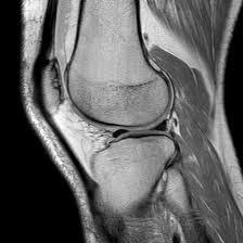
 The Magnetic resonance is an imaging study widely used today for diagnostic purposes, since it allows the soft tissue visualization that cannot be seen on plain radiographs or by others media.
In this technique dating from the eighties, you get images details of any part of the body by means of app of magnetic fields with powerful circular magnets that cause the nuclei of hydrogen atoms found throughout the body to align and absorb EnergyOnce this field ceases, these particles radiate the energy that is captured by a receiving antenna, thus originating the different images, shown as cuts made approximately every 5 millimeters along the structure to be studied.
In cases where for some reason a better visualization of a structure is required, a paramagnetic contrast medium called Gadolinium, which is applied intravenously before taking the images. This contributes to increasing the intensity signal of some lesions, especially infections, tumors and metastases.
The MRI study can be done in any part of the body, however it has a greater use in the cases detailed below:
1. When a lesion of the intervertebral discs is suspected, as in the case of herniated discs, where this study is able to show the lesions that the discs and the degree of compromise on the nerve roots and the spinal cord spinal.
2. In the case of a cerebrovascular accident, here the MRI allows to identify if it is due to thrombosis or hemorrhage and even the degree of involvement of the different structures of nervous system and possible complications such as hydrocephalus.
3. To confirm the presence of some types of tumors, since it allows a better visualization of the different organs, revealing abnormal structures that may be compatible with injuries malignant.
4. In order to carry out the determination of the stage of a cancer, by allowing to evaluate if there is involvement of organs such as the lymph nodes, the liver, the lungs or the brain.
5. Visualization of vascular structures such as the arterial or venous system, using the technique known as magnetic resonance angiography, especially useful in diagnosing blocked arteries coronary.
6. For him diagnosis of non-tumor lesions of the nervous system such as infections, arteriovenous malformations, aneurysms, cysts, and demyelinating diseases such as multiple sclerosis and lateral sclerosis amyotrophic.
 In this type of study, no type of radiation is originated, since it is based on the application of magnetic fields. Although it is a safe and non-invasive study, MRI cannot be performed in the case of people who have metallic elements in their body since these can be attracted towards the magnet moving from the site where they are located causing injuries. In the same way, it cannot be applied to people with implants of electronic devices such as pacemakers or hearing aids in the inner ear.
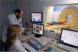
It is possible that during this study patients feel somewhat anxious as they must be introduced into a narrow tunnel that is nothing more than the cavity of a Large magnet, in the case of people who suffer from claustrophobia it is usually necessary to practice some type of sedation to be able to undergo this procedure, the latter has led to the development of open resonators where the patient is subjected to the magnetic field without the need to be inside a tunnel.
Topics in Magnetic Resonance


